
The permanent dentition is made up of 32 teeth, and each tooth serves a unique purpose in the mouth. Of these 32 teeth, four are the longest: the upper and lower central incisors, and the upper and lower first molars. The length of these teeth varies from person to person, but they are generally longer than other permanent teeth in the mouth. This article will discuss which teeth are the longest in the permanent dentition and why they are important for proper oral health.The longest teeth in the permanent dentition are the maxillary and mandibular central incisors. These teeth are located at the center of the jaw, top and bottom, and measure up to 22mm in length.
Anatomy Of The Permanent Dentition
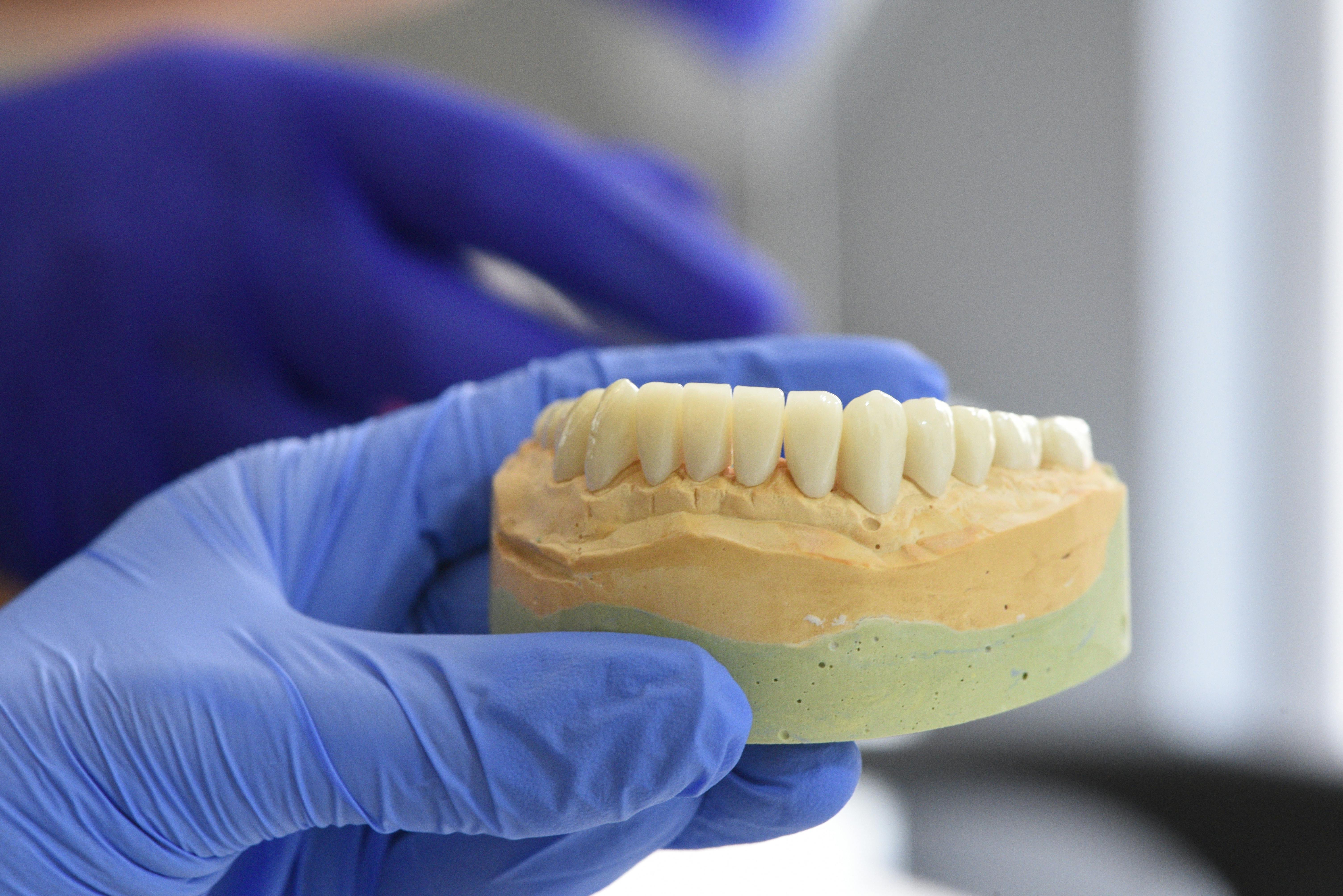
The anatomy of the permanent dentition is a complex subject. It involves understanding the structure of the teeth, their arrangement in the mouth, and their function. The permanent dentition consists of three major types of teeth: incisors, canines, and molars. Each type of tooth is composed of different parts that are important for proper functioning.
The incisors are located at the front of the mouth and are used for cutting or tearing food. They consist of a crown, root, enamel, dentin, and pulp chamber. The crown is the visible portion above the gum line and contains enamel which protects the underlying layers from decay or trauma. The root is located below the gum line and anchors the tooth into its socket in the jawbone. The dentin is found beneath the enamel and contains a pulp chamber which houses nerve endings that provide sensation to the tooth when it comes in contact with food or other substances.
The canines are located on either side of each incisor and are used for ripping or tearing food apart before it is swallowed. They consist of a sharp crown with a single root that helps to anchor it firmly into its socket in the jawbone. Canines also contain enamel on their crowns to protect them from decay or trauma as well as dentin underneath which houses a pulp chamber containing nerve endings that help provide sensation to them when they come into contact with food or other substances.
The molars are located towards the back of each side of the mouth and are used for grinding up food before it is swallowed. They consist of several cusps on their crowns which allow them to better chew up food before it is swallowed as well as roots that help to anchor them securely into their sockets in the jawbone. Molars also contain enamel on their crowns to protect them from decay or trauma as well as dentin underneath which houses a pulp chamber containing nerve endings that help provide sensation to them when they come into contact with food or other substances.
Overall, understanding anatomy of permanent dentition helps us understand how our teeth work together to properly break down our food so we can swallow it more easily without any problems. It also helps us identify any potential issues such as decay or trauma before they become serious problems that require more extensive dental care treatments such as braces or fillings.
Size Of The Permanent Teeth
Permanent teeth are larger than baby teeth, and they usually come in around age 6. The size of the permanent teeth is determined by the size of the jawbone, and this can vary from person to person. Permanent teeth are usually wider than baby teeth, and they may also be longer. They are also more likely to have ridges or furrows on them, which gives them a distinctive appearance. Permanent teeth typically have a more yellowish hue than baby teeth due to the enamel being thicker.
The size of your permanent teeth can also be affected by genetics, or by any damage that has been done to your baby teeth prior to the eruption of the permanent ones. If a baby tooth is lost too soon or damaged, then the permanent tooth will not have enough room to erupt properly and may be smaller than it should be. This can lead to crowding in the mouth as well as other dental issues that need to be addressed in order for your oral health to stay in good condition.
It is important that you visit your dentist regularly so that any potential problems with your teeth can be identified and treated early on before they become more serious. Your dentist will be able to give you advice on how to take care of your permanent teeth so that they stay healthy and strong for years to come.
Primary Dentition
Primary dentition refers to the first set of teeth that a child has. It consists of 20 teeth, including 10 incisors, four canines, and six molars. Primary dentition begins to appear in the mouth at around 6 months of age and is typically complete by age 3. As primary teeth erupt into the mouth, they help guide the permanent teeth into their correct positions. Primary teeth are also important for helping children learn how to speak and chew properly. They also help maintain space in the jaw for permanent teeth to come in.
Permanent Dentition
Permanent dentition refers to the second set of adult teeth that replace the primary dentition over time. This set consists of 32 permanent teeth, including 12 incisors, four canines, eight premolars, and eight molars. Permanent dentition typically begins erupting around age 6 and is usually complete by age 21. Permanent dentition is often referred to as adult teeth because they are intended to last for life if taken care of properly. Unlike primary dentition, which is made up of mostly baby-like teeth with rounded edges and small crowns, permanent dentition is made up of larger and flatter adult-like molars with sharper edges that are better suited for grinding food.
The Longest Teeth in the Permanent Dentition
The longest teeth in the permanent dentition are the maxillary and mandibular first molars, which are located at the back of the mouth on both sides of the jaw. They are typically the biggest and strongest teeth in both the upper and lower jaws. The maxillary first molars have four cusps, while the mandibular first molars typically have five cusps. These teeth have broad surfaces that help them to grind food and they are used for chewing.
Maxillary first molars are usually larger than mandibular first molars, though this can vary from one person to another. They also have a longer root compared to other teeth in both upper and lower jaws, with an average length of up to 25mm. The root of a mandibular first molar is usually shorter, averaging around 20mm in length.
The maxillary and mandibular first molars play an important role in occlusion (or bite) since they meet each other when the mouth is closed. They also help to maintain balance between opposing forces while chewing, as well as help support facial structures such as cheeks and lips. As such, it is important that these teeth remain healthy and strong so that they can perform their function effectively.
Regular visits to a dentist are necessary for maintaining good oral health, including keeping long-term dental conditions such as cavities or gum disease at bay. During routine check-ups, dentists may identify any potential issues with the longest teeth in the permanent dentition before they become more serious problems by carrying out examinations or taking x-rays of these areas. If any issues are identified, appropriate treatments can be recommended by dentists to prevent further damage or deterioration of these important teeth.
Mandibular Canines: The Longest Teeth in Permanent Dentition
The mandibular canines, also known as the lower canines, are the longest teeth in the permanent dentition. These teeth are situated between the lateral incisors and premolars in both jaws and have an average length of about 25 millimeters. They are usually pointed or curved in shape and are used for tearing food. They are also referred to as ‘cuspids’ or ‘eye teeth.’
The mandibular canines are made up of two cusps, a mesial (toward the midline) and a distal (away from the midline). These cusps allow for a greater surface area for chewing on tough foods such as meat. The crowns of these teeth have a broad base with distinct ridges which aids in gripping food. The roots of these teeth usually have two roots with an average length of 12-14mm each.
In comparison to other types of teeth, mandibular canines have a hard enamel layer that protects them from wear and tear due to their location within the mouth which is constantly exposed to acidic foods and beverages. This enamel layer also helps them maintain their pointed shape over time. Mandibular canines require regular brushing and flossing to keep them healthy and free from plaque buildup which could lead to tooth decay or gum disease.
Overall, mandibular canines play an important role in helping us chew our food and maintaining our oral health. It is important to take good care of these teeth by brushing twice daily, flossing regularly, eating healthy foods, avoiding sugary snacks, rinsing with mouthwash after meals and visiting your dentist at least twice a year for regular check-ups.
Maxillary Canines: Second Longest Teeth In Permanent Dentition
Maxillary canines, also known as cuspids, are the second longest teeth in permanent dentition. They are located between the lateral incisors and first premolars in the upper jaw. Maxillary canines have a distinct shape with sharp points and a large, flat surface on their biting edge. They have two cusps on their biting surface which help them tear food apart during chewing. The roots of maxillary canines are usually longer than other teeth and they typically grow in an upright position. Maxillary canines play an important role in maintaining proper occlusion and are often used as an anchoring point for orthodontic treatment. In addition, maxillary canines help to support the lips and cheeks to prevent sagging or wrinkling of the skin around the mouth.
Maxillary canines are also one of the most difficult teeth to restore due to their unique shape and position. Because of their size and shape, it is important that they be restored properly so that they do not interfere with other teeth or impair function. If maxillary canines become decayed or damaged, they may need to be removed and replaced with a dental bridge or implant to restore proper function and aesthetics.
Overall, maxillary canines are essential for proper bite alignment, supporting facial tissues as well as providing structure for orthodontic treatment. It is important that they remain healthy and free from decay or damage in order to maintain proper oral health and function. Regular visits to the dentist for checkups, cleanings, and restorative treatment can help keep maxillary canines healthy for many years to come.
Enamel Thickness Of Mandibular Canines
Enamel thickness of mandibular canines is an important factor when studying the development and growth of the teeth. It is important to measure the enamel thickness of the mandibular canines in order to determine if there is any abnormality or pathology present. The enamel of the mandibular canines is generally thicker than that of other teeth, making it easier to measure accurately. This allows for a more accurate assessment of the morphology and structural integrity of the tooth.
The enamel thickness of mandibular canines will vary depending on a number of factors including age, genetics, diet, and lifestyle. As a person ages, their teeth may become more brittle due to wear and tear, leading to an increase in enamel thickness. Genetics may also play a role in enamel thickness, as certain genetic traits may lead to thicker or thinner enamel than usual. Diet and lifestyle may also affect how much enamel is produced by the body; for example, those who drink acidic beverages on a regular basis may have thinner than average enamel on their teeth because acids erode away at tooth enamel over time.
When measuring the enamel thickness of mandibular canines, dental professionals typically use an imaging technology such as dental radiographs (x-rays) or computed tomography (CT scans). These technologies allow for precise measurements that are accurate within fractions of millimeters. By measuring this information accurately, dentists are able to identify any abnormalities or pathology related to the teeth structure that could be causing pain or discomfort to patients.
It is important for dentists to assess the enamel thickness of mandibular canines in order to provide accurate diagnosis and treatment plans for their patients. Accurate measurements allow dentists to properly detect any underlying issues with tooth structure and provide effective treatments accordingly. With proper measurements and analysis, patients can receive high-quality care that will help them maintain healthy teeth for many years into the future.

Conclusion
The longest teeth in the permanent dentition are the maxillary and mandibular first molars, which can measure up to three-quarters of an inch in length. While they are not the only teeth that can be considered long, they are certainly the longest ones. The other teeth in the permanent dentition may be slightly longer or shorter than average, but none of them come close to matching the size of the first molars. It is important to take good care of all your teeth, regardless of their length, since any tooth decay could lead to a host of dental problems. With proper oral hygiene and regular visits to a dentist, you can keep your teeth healthy for as long as possible.
In conclusion, the longest teeth in your permanent dentition are your maxillary and mandibular first molars. While their size may vary from individual to individual, these two teeth tend to be significantly longer than any other type of tooth in the mouth. Taking good care of all your teeth is essential for maintaining good oral health.