
Reading a teeth x-ray can provide an oral health professional with valuable insight into the condition of your teeth and jaw structure. A teeth x-ray is a form of diagnostic imaging that uses a low dose of radiation to capture an image of the inside of your mouth. By looking at the x-ray, your dentist or orthodontist can diagnose problems such as cavities, impacted teeth, bone loss, gum disease, and even tumors. In this article, we’ll discuss how to read a teeth x-ray so you can have a better understanding of what your dentist is looking for when they examine the results.Understanding teeth X-ray images can help dentists properly diagnose and treat dental conditions. X-rays provide a detailed view of the inner structures of the teeth, including cavities, periodontal disease, tooth decay and bone loss. X-rays also help dentists detect changes in existing dental work, such as crowns or fillings, as well as any abnormalities or trauma to the tooth’s roots and surrounding tissues. By examining an X-ray image of a patient’s teeth, a dentist can more accurately assess and diagnose the condition of their teeth.
Preparing for a Teeth X-Ray
Having a teeth x-ray is an important part of preventive dental care. X-rays help your dentist detect any problems that may not be visible during a regular dental exam. Before having a teeth x-ray, it’s important to prepare properly to ensure accurate results.
It’s best to arrive at your appointment with clean teeth, so make sure you brush and floss thoroughly before the appointment. It’s also important to remove any jewelry you are wearing, as it can interfere with the x-ray images. You may also need to remove dentures or other dental appliances before the procedure.
Your dentist will provide you with a lead apron to protect your body from radiation exposure during the x-ray. It is important that you wear this apron as directed for the duration of the procedure. In addition, your dentist may provide you with protective eyewear or ask you to close your eyes during the x-ray process.
Before taking an x-ray, your dentist will ask questions about medical history and any medications that you may be taking, as some medications can interfere with results. Additionally, if you are pregnant or think there is a chance that you may be pregnant, let your dentist know prior to having an x-ray taken.
By following these preparations steps before having a teeth x-ray taken, you can help ensure accurate results and reduce radiation exposure while giving your dentist valuable information about your oral health condition.
Types of Teeth X-Rays
X-rays are an important tool for diagnosing and treating dental problems. They allow dentists to see what is happening below the surface of the teeth and gums. There are several types of X-rays that can be used to diagnose and treat dental issues. The most common types of X-rays are bitewing, periapical, occlusal, and panoramic.
Bitewing X-Rays
Bitewing X-rays are usually taken annually at a patient’s regular checkup. These images provide a view of the upper and lower back teeth on one side of the mouth. They are typically used to identify cavities, tartar buildup, and bone loss due to periodontal disease.
Periapical X-Rays
Periapical X-rays provide a detailed view of one or two teeth at a time including the entire tooth structure from crown to root tip. This type of X-ray is usually taken when there is an issue with a particular tooth such as pain or swelling. It can also be used to assess impacted wisdom teeth before they are removed or evaluate possible root canal treatments.
Occlusal X-Rays
Occlusal X-rays show both arches in the mouth in one image which makes it easier for dentists to evaluate how the upper and lower teeth fit together when biting down (occlusion). This type of imaging is often used in orthodontic treatment planning when braces are being considered.
Panoramic X-Rays
Panoramic X-rays provide a wide angle overview of all teeth including those that may be impacted or difficult to see with other types of imaging. These images typically include the jawbone, sinuses, nasal area, and other surrounding structures which makes them useful for assessing facial trauma or diagnosing temporomandibular joint (TMJ) disorders.
What Are the Uses of Teeth X-Rays?
Teeth X-rays are an important diagnostic tool used by dentists to help identify problems related to the teeth and surrounding structures. X-rays are used to detect cavities, bone loss, abscesses, cysts, impacted teeth, and other abnormalities in the mouth. They also provide information about the relationship between the upper and lower jaws. X-rays can also be used to evaluate the growth and development of a patient’s teeth.
X-rays can be used to help diagnose a wide range of dental conditions such as caries, periodontal disease, tooth decay, abnormalities in jaw structure and development, impacted teeth, abscesses and bone loss. By using X-ray images dentists can see things that they cannot see with just a visual examination. This makes it easier for them to diagnose problems quickly and accurately.
X-rays are also useful for planning orthodontic treatments such as braces or Invisalign aligners. The X-ray images are also used to plan dental implant procedures or bridge placements. They can be used to assess periodontal disease by looking for signs of bone loss around the roots of teeth or infection in the gums.
In addition to diagnosing problems with teeth and gums, X-rays can also be useful for detecting tumors or other abnormal growths in the mouth. X-ray images can help identify areas where surgery may be necessary in order to remove tumors or abnormal tissue masses from the mouth.
Overall, teeth X-rays play an important role in helping dentists accurately diagnose various dental conditions and plan treatments accordingly. They provide important information about both visible and hidden problems that may not be detected during a visual examination alone.
What Can You Learn from a Teeth X-Ray?
X-rays of the teeth can provide valuable insight into the health of your oral cavity. They can help your dentist identify problems such as cavities, bone loss, impacted teeth, and other issues that cannot be seen with the naked eye. X-rays may also be used to diagnose periodontal disease and to plan orthodontic treatment.
X-rays of the teeth allow dentists to detect cavities in between teeth, which can be difficult to spot with a visual exam alone. They can also help determine how much decay is present in a tooth and if it has spread into the root or surrounding bone. Additionally, they can detect any abnormalities such as cysts or tumors in the jawbone.
X-rays are also used to evaluate the growth and development of the teeth in children. These images show if each tooth is erupting properly and at what rate it is growing. X-rays can also reveal impacted teeth or those that are coming in at an incorrect angle.
In addition to helping diagnose oral health issues, x-rays are important for making treatment decisions for orthodontic care. They give dentists a better understanding of the position of each tooth so that they can develop an effective treatment plan.
Overall, dental x-rays provide valuable information about a person’s oral health that cannot be gleaned from a visual exam alone. They allow dentists to identify problems early on and develop effective treatment plans for their patients.
What Do Different Colors on a Teeth X-Ray Mean?
A dental x-ray is an important tool used by dentists to diagnose and treat various dental conditions. Dental x-rays use different colors to represent different types of tissue in the patient’s mouth. The colors can help dentists determine the presence of cavities, infection, impacted teeth, and other problems.
The most common color used in a dental x-ray is black. Black represents hard tissue such as enamel and dentin, which are both components of teeth. White is also often used to represent soft tissue like gums and bone. Grey is commonly used to represent areas that are not healthy or do not contain any tissue at all, such as abscesses or areas that have been filled.
Other colors may be seen on a dental x-ray depending on the type of imaging system being used. Different imaging systems use different color schemes for different types of tissues; for example, some systems use blue to show bone while others may use yellow instead. Additionally, certain imaging systems may add extra colors such as pink or green to indicate specific types of abnormalities or conditions.
It is important for dentists to be able to interpret the various colors on a dental x-ray accurately in order to properly diagnose and treat their patients’ conditions. By understanding what each color means, dentists can gain valuable insight into their patients’ mouths and help them maintain good oral health.
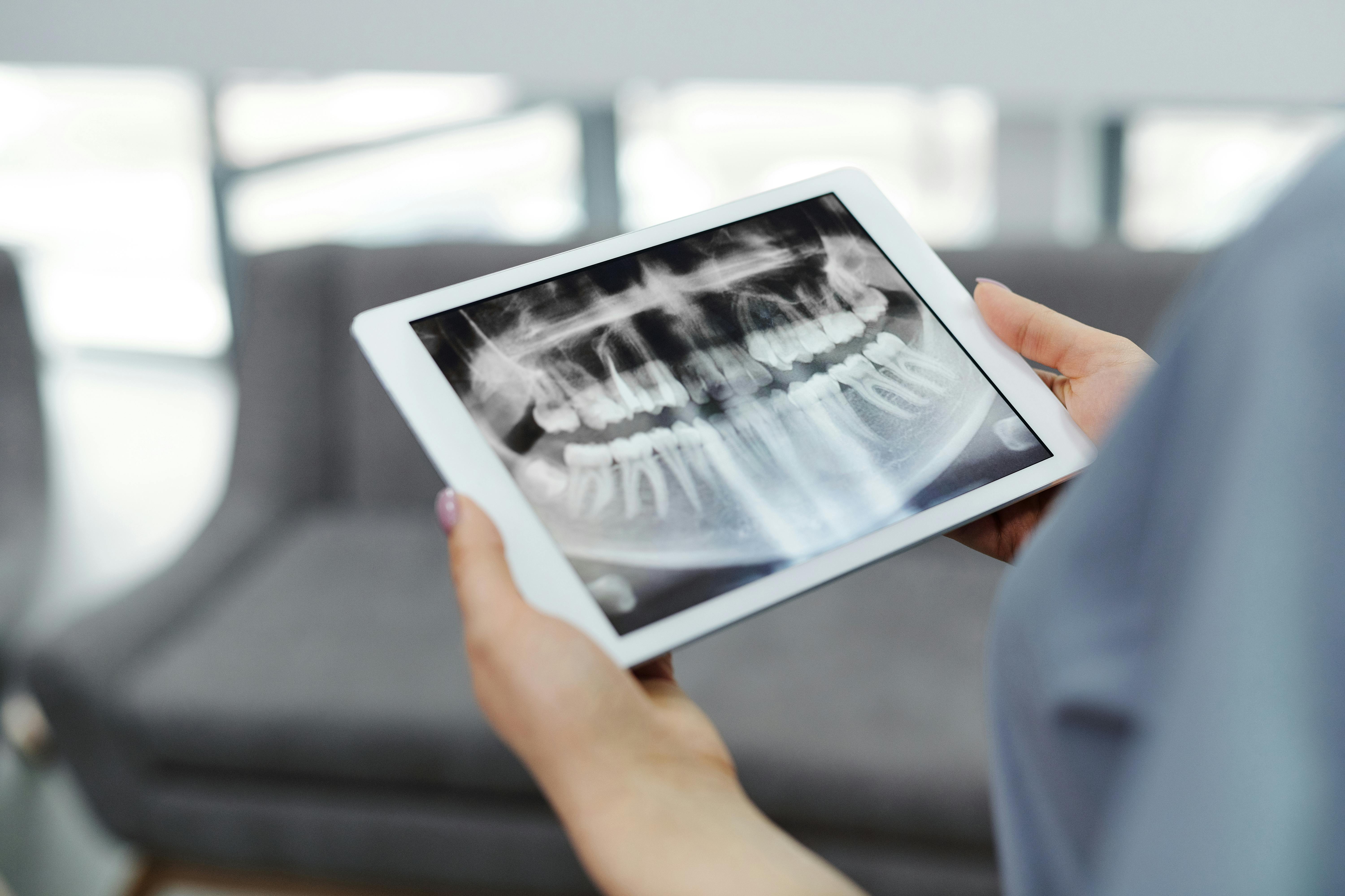
Interpreting a Teeth X-Ray Image
Interpreting a teeth x-ray image is a crucial part of diagnosing various types of dental problems. X-rays provide detailed images that allow dentists to see what is happening inside the teeth and mouth. By looking at an x-ray image, dentists can diagnose cavities, impacted teeth, gum disease, and other issues that may be present in the mouth. In order to interpret the image correctly, it is important to understand the different parts of a teeth x-ray image and how they relate to each other.
The first thing to look for when interpreting an x-ray image is the size and shape of the teeth. The size of each individual tooth can tell dentists a lot about the overall health of the mouth. For example, smaller than average teeth may indicate that there may be some bone loss due to periodontal disease or other oral health issues. The shape of the teeth can also provide clues about any problems in the mouth such as overcrowding or misalignment.
The second aspect to consider when interpreting an x-ray image is the condition of the supporting structures such as gums and jawbone. These structures provide support for the teeth and if they are not healthy, then it could mean that there could be potential problems with dental health down the line. Gingivitis and periodontal disease are two common issues that can be diagnosed by looking at an x-ray image. The amount of bone loss can also be determined by examining an x-ray image which can help dentists decide on treatment options for their patients.
Finally, it is also important to look at any abnormalities in an x-ray image such as cysts or tumors which may indicate underlying dental issues such as infection or trauma. Any changes in color or texture should also be noted as they could be indicative of certain types of conditions such as cavities or decay. By analyzing all these aspects together, dentists can make an accurate diagnosis based on what they see on an x-ray image.
Common Findings on Teeth X-Rays
X-rays of teeth are commonly used by dentists to detect and diagnose dental issues. X-rays provide a view of the inside of the teeth, allowing dentists to see abnormalities and conditions that may not be visible to the naked eye. Through x-ray imaging, dentists can identify cavities, impacted teeth, and other issues that may require treatment. Additionally, x-rays can be used to monitor the progress of dental treatments, such as braces or root canals.
Some common findings on teeth x-rays include cavities or lesions in the enamel, signs of infection or decay in the jawbone, impacted teeth (teeth that have not emerged from the gums), damage to bones surrounding the roots of a tooth, cysts or tumors in the jawbone, and bone loss due to gum disease. In addition to these findings, x-rays may also reveal abscessed teeth, which are caused by bacterial infections around a tooth’s root. X-ray images can also provide information about wisdom teeth development and presence of plaque buildup.
X-rays can also be used to detect signs of orthodontic problems such as crowding or misalignment of teeth. Furthermore, they may show signs of temporomandibular joint (TMJ) disorder caused by bruxism (teeth grinding). In some cases, x-rays may even reveal tumors in the mouth or jawbone that require further evaluation and treatment.
In conclusion, x rays are an important diagnostic tool for dentists as they allow them to accurately diagnose a variety of dental issues and conditions. Common findings on teeth x-ray images include cavities or lesions in enamel, impacted teeth, cysts or tumors in the jawbone, bone loss due to gum disease and abscessed teeth. X rays can also be used to detect signs of orthodontic problems such as crowding or misalignment and TMJ disorder caused by bruxism.

Conclusion
Reading a teeth X-ray is a skill that requires patience and practice. It is necessary to understand the anatomy of the teeth as well as the basics of radiography for accurate interpretation of the images. With regular practice, one can improve their skills to interpret teeth X-rays accurately and effectively. Furthermore, an understanding of dental disease processes can also aid in diagnosis.
In conclusion, it is essential to remember that reading a teeth X-ray is not a simple task and requires dedicated practice in order to become proficient. With the right approach and dedication, one can become an expert in interpreting teeth X-rays and make accurate diagnoses for better patient outcomes.